 Dachshunds are significantly overrepresented when it comes to IVDD, and this is largely due to their genetic chondrodystrophic (dwarf) makeup, which causes premature disc degeneration.
The primary genetic factor involved is an FGF4 retrogene insertion on Chromosome 12 (CFA12), which leads to premature degeneration and calcification of the nucleus pulposus — the inner jelly-like material of the disc.
When this is paired with an earlier mutation on Chromosome 18 (CFA18), which contributes to the Dachshund’s characteristic short-legged build, the combination results in a significantly higher risk of IVDD.
This is why many countries place a strong emphasis on early recognition and responsible breeding practices.
Radiographic screening of the spine between 24 and 48 months of age can help detect calcified discs. Dogs showing a high number of calcified discs are often excluded from breeding programs.
DNA risk screening is also available, although it is not always considered a fully reliable standalone tool.
The key takeaway here is to research breeders carefully and choose those who support ethical breeding programs aimed at reducing the prevalence of IVDD.
Here comes a little anatomy lesson.
Let’s first look at a side view of the T13, L1 and L2 vertebrae to get a general bearing of anatomy and a view of where the discs and spinal cord sit in relation to each other.
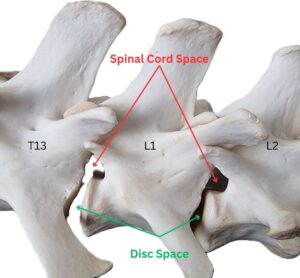
Intervertebral discs sit between each vertebra in the spine. Their main role is to provide cushioning and shock absorption.
Each disc is made up of: 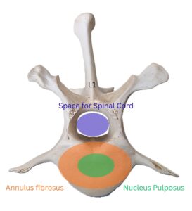
- an outer fibrous ring (annulus fibrosus)
- an inner gelatinous centre (nucleus pulposus)
The spinal cord runs directly above these discs, extending from the brain all the way to the tail.
The easiest way I explain IVDD to owners is to imagine a jam donut.
The donut itself is the disc, and the jam inside represents the nucleus pulposus.
In Dachshunds, this inner jelly-like material often calcifies and hardens much earlier in life.
Instead of remaining soft and shock-absorbing, it becomes brittle.
Then, after something as simple as:
- jumping off the couch
- twisting sharply
- slipping on smooth floors
- rough play
- or simply gradual degeneration over time
…the hardened disc material can suddenly burst through the outer fibres and compress the spinal cord.
This compression is what causes the neurological signs we see.
The severity depends on:
- how much disc material extrudes (volume)
- how quickly it happens (velocity)
- the force of compression (volume and velocity), which determines the amount of bruising or trauma within the spinal cord
To help visualise this, imagine two different scenarios:
Being hit by a bike travelling at 10 km/h is very different from being hit by a truck travelling at 100 km/h. The outcomes would be vastly different.
The same applies to the spinal cord — the greater the force, the greater the trauma and bruising.
IVDD is generally graded from 1 to 5 to measure the severity of spinal cord compression and injury.
This grading system is critical for determining:
- prognosis
- treatment options and likely outcome
- monitoring deterioration after the IVDD injury
- monitoring recovery after treatment
Grade 1: Pain only, still walking normally
Grade 2: Weakness and wobbliness but still walking (Ambulatory Paraparesis)
Grade 3: Unable to walk independently but still showing some voluntary movement (Non-ambulatory Paraparesis)
Grade 4: No voluntary hind limb movement, but deep pain sensation is still present (Paraplegia)
Grade 5: No movement and loss of deep pain sensation (Paraplegia with loss of deep pain perception)
This grading system is one of the most important factors in deciding whether surgery or conservative management is recommended.
Usually, your GP vet will be the first point of contact.
They will begin with a full neurological examination, checking:
- reflexes
- limb strength
- proprioception
- pain sensation
- ability to stand and walk
This initial neurological assessment helps determine the severity of the issue and guides the next steps.
With Grade 1 or 2, your vet may recommend:
- strict crate rest
- pain relief and anti-inflammatory
- close monitoring
or they may refer you directly to a surgical specialist for further diagnostics and possible surgery.
To confirm the exact location and severity, advanced imaging is usually required and will either be done via MRI or CT with a myelogram.
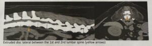
Both methods allow the surgeon to identify:
- the exact location of spinal cord compression
- the extent of disc material extrusion
- whether the compression is lateralised to one side
This information is essential for planning the surgical approach and ensuring all compressive disc material is removed.